What Is CariVu?
CariVu is a handheld diagnostic device made by DEXIS that shines near-infrared light through a tooth to create an image of its internal structure. The device helps dentists spot cavities, cracks, and other problems hidden inside the tooth.
Traditional dental X-rays use ionizing radiation to produce images of teeth. CariVu takes a different approach. It uses near-infrared transillumination, often shortened to NIRT. This means it passes a specific wavelength of light through the tooth. Healthy tooth structure allows the light to pass through evenly. Areas of decay, fractures, or damage absorb or scatter the light differently, showing up as dark spots on the resulting image.
The images CariVu produces look similar to a standard bitewing X-ray. Dentists can read them using the same visual logic they already know. Because the technology involves light rather than ionizing radiation, a dentist can take as many images as needed during a single visit. This makes it especially useful when repeated imaging is needed to monitor a developing area of concern. For context, modern dental X-rays also deliver very low radiation doses. A set of four digital bitewing X-rays delivers approximately 0.005 mSv, which is less than a single day of natural background radiation. [2] [3]
CariVu is used in general dentistry and by specialists, including endodontists who focus on diagnosing and treating problems inside the tooth. You can learn more about what these specialists do on the endodontics page.
How CariVu Works: The Technology Explained
CariVu passes near-infrared light through a tooth, and a sensor on the opposite side captures the light that comes through to create a diagnostic image.
Near-Infrared Transillumination (NIRT)
The device uses light in the near-infrared spectrum, typically at a wavelength of approximately 780 nanometers. This is just beyond the range of visible light. At this wavelength, healthy enamel (the hard outer shell of a tooth) is largely transparent. Light passes through it relatively easily.
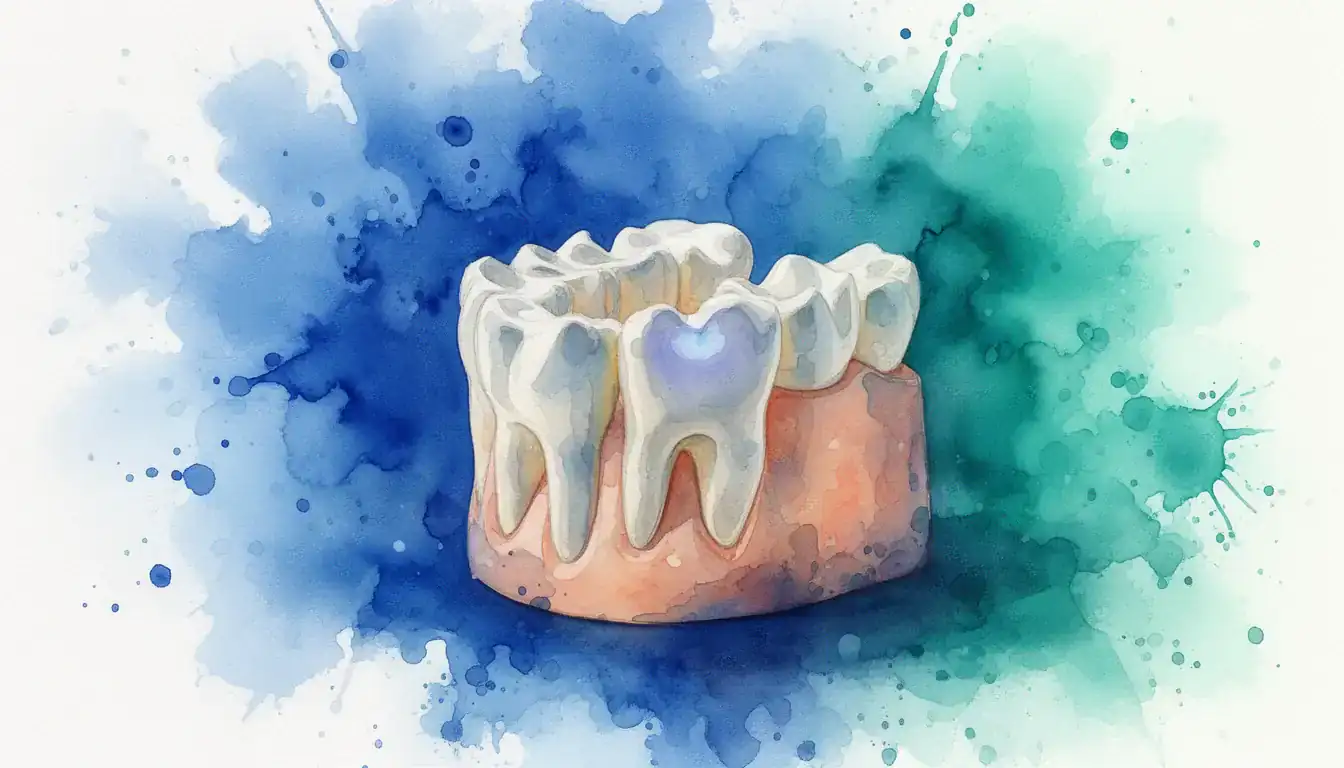
When the light hits an area of decay or a crack, the damaged structure changes how the light travels. Decay absorbs more of the near-infrared energy. Cracks scatter and redirect it. The sensor on the other side of the tooth records these differences. Software then converts the data into a grayscale image that looks much like a dental X-ray.
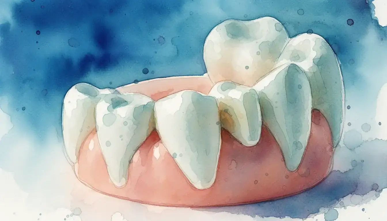
The key principle is contrast. Healthy enamel appears bright or translucent on the image. Decayed areas or fractures appear as dark shadows. Dentin (the softer layer beneath the enamel) appears slightly different from enamel, allowing dentists to estimate how deep a problem might extend.
What the Imaging Process Looks Like
The CariVu device looks like a small, curved clip that fits over the crown of the tooth. Two soft, flexible pads rest against the sides of the tooth. One side emits the near-infrared light. The other side contains the sensor.
During the scan, the room lights are typically dimmed. The pads gently compress against the tooth to block ambient light. The entire process for a single tooth takes only a few seconds. The image appears on a computer screen in the operatory almost immediately.
No lead apron is needed. No thyroid collar is needed. The patient simply sits in the dental chair while the clinician places the device around each tooth being examined.
When Dentists Use CariVu
CariVu is primarily used to detect interproximal caries (cavities between teeth), but it also has applications in identifying cracks and monitoring lesions over time.
Cavity Detection Between Teeth
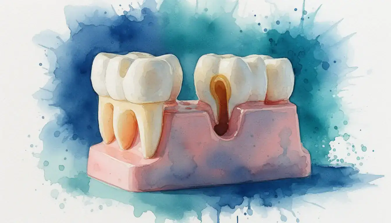
The most common use for CariVu is finding cavities on the surfaces where two teeth touch each other. These are called interproximal surfaces. Cavities in these areas are difficult to see during a visual exam because the teeth block the view. Traditionally, bitewing X-rays are the standard tool for finding them. [2]
CariVu provides an alternative way to image these hidden areas. When a cavity is present between two teeth, the decayed enamel absorbs the near-infrared light. It shows up as a dark shadow on the image. This can help confirm a diagnosis when an X-ray finding is unclear, or it can serve as a radiation-free screening option.
Identifying Cracks and Fractures
Cracks in teeth are notoriously difficult to diagnose. They often do not appear on standard dental X-rays. An endodontist evaluating a tooth for a possible crack may use transillumination as part of the diagnostic process. [1]
When near-infrared light encounters a crack, the fracture line disrupts the path of the light. The crack appears as a distinct dark line on the image. This can help an endodontist determine whether a crack is present, where it is located, and how it relates to the patient's symptoms. CariVu is one of several transillumination tools that may be used for this purpose.
Monitoring Early Decay Over Time
Not every spot of early decay requires a filling. Sometimes a dentist will choose to monitor a small area of demineralization (the earliest stage of a cavity) to see if it progresses or stabilizes. Because CariVu involves no ionizing radiation, images can be taken at every visit without concern for cumulative exposure.
This makes it a practical tool for tracking early lesions. The dentist can compare images from one visit to the next to see if the dark area has grown, stayed the same, or improved with preventive treatments like fluoride.
Use with Children and Pregnant Patients
Children and pregnant patients are populations where providers are especially mindful about imaging decisions. Modern dental X-rays deliver very low doses of radiation and are considered safe for these groups when clinically necessary. A set of four digital bitewing X-rays exposes a patient to roughly 0.005 mSv, which is less than the approximately 0.01 mSv a person receives from a single day of natural background radiation. [3] Still, some patients and providers prefer a radiation-free option when the clinical question can be answered with one. [2]
CariVu can be a useful option in these situations. It allows the clinician to gather diagnostic information about potential cavities without any ionizing radiation. However, it does not replace the full diagnostic scope of X-rays, so the decision to use CariVu alone or alongside X-rays depends on the specific clinical situation. Patients should feel comfortable knowing that both CariVu and properly indicated dental X-rays are safe tools.
Evidence and FDA Status
CariVu has received FDA 510(k) clearance as a diagnostic aid, which means the FDA determined it is substantially equivalent to an existing legally marketed device for its intended purpose.
FDA Clearance vs. FDA Approval
It is important to understand the difference between FDA clearance and FDA approval. FDA clearance, granted through the 510(k) pathway, means the device has been reviewed and found to be substantially equivalent in safety and effectiveness to a device already on the market. FDA approval, on the other hand, involves a more rigorous premarket approval (PMA) process typically reserved for higher-risk devices.
CariVu has received FDA 510(k) clearance. This is the standard regulatory pathway for most dental diagnostic devices. It confirms that the device meets safety and performance criteria for its labeled use as an aid in caries detection.
What Research Shows
Several published studies have evaluated near-infrared transillumination technology for caries detection. Early research suggests that NIRT performs comparably to bitewing radiographs for detecting interproximal caries in enamel, though results vary across different study designs and tooth types.
Some studies indicate that NIRT may have high specificity, meaning it is good at correctly identifying teeth that do not have cavities. Its sensitivity, the ability to correctly identify teeth that do have cavities, has varied more widely across studies. This means CariVu may be better at confirming that a tooth is healthy than at catching every instance of early decay.
Research on NIRT for crack detection is more limited. Transillumination in general has long been used by endodontists as a clinical tool for identifying cracks, and CariVu represents a more standardized, digital version of this technique. [1] More research with larger sample sizes and standardized methods would strengthen the evidence base.
Professional Organization Perspectives
The American Dental Association supports the use of evidence-based diagnostic tools and emphasizes that any imaging method should be selected based on the individual patient's clinical needs. [2] The American Association of Endodontists recognizes transillumination as part of the diagnostic toolkit for evaluating cracked teeth and other endodontic conditions. [1]
Neither organization has issued a specific position paper endorsing or opposing CariVu by name. The general consensus in the dental profession is that NIRT devices like CariVu are useful supplementary tools rather than standalone replacements for radiographic imaging.
Benefits and Limitations of CariVu
CariVu offers clear advantages in specific situations but also has limitations that patients and providers should understand.
Advantages
One key benefit is the absence of ionizing radiation. Every CariVu image is radiation-free. This is especially useful when frequent monitoring is needed, such as tracking a suspicious area over several visits. It also provides a practical alternative for children and pregnant patients, though it is worth noting that modern dental X-rays deliver very low radiation doses and are also considered safe for these groups when clinically indicated. [2] [3]
The images are generated instantly and displayed on a chairside monitor. This allows the dentist to show you exactly what they see in real time. Many patients find it easier to understand a CariVu image compared to a traditional X-ray because the decay appears as a clear dark area against a bright, healthy tooth structure.
Because there is no ionizing radiation, there are no regulatory limits on how many images can be taken. A dentist can image every tooth in the mouth during a single visit if the clinical situation calls for it. Repeat imaging at follow-up visits carries no cumulative exposure concern.
- No ionizing radiation exposure, allowing unlimited repeat imaging
- Images produced in seconds with no processing time
- No lead apron or thyroid collar required
- Practical for frequent monitoring of early lesions over multiple visits
- Helpful for patients who are anxious about X-rays
- Real-time images that are easy for patients to understand
Limitations
CariVu cannot image below the gumline. It works by passing light through the crown of the tooth. This means it cannot evaluate bone levels, root infections, or conditions deep within the jawbone. Standard X-rays and cone-beam computed tomography (CBCT) scans remain necessary for those diagnoses. [1]
The device works best on natural tooth structure. Teeth with large existing restorations (fillings, crowns, or veneers) can be difficult to image accurately because the restorative materials block or alter the near-infrared light in unpredictable ways.
Sensitivity for very early enamel lesions has varied in studies. A tooth that appears clear on CariVu may still have a very small area of early demineralization that the device cannot resolve. For this reason, most clinicians use CariVu alongside X-rays rather than in place of them.
- Cannot image roots, bone, or structures below the gumline
- Less effective on teeth with large existing restorations
- Variable sensitivity for the earliest stages of decay
- Does not replace the full diagnostic scope of dental X-rays
- Not all dental offices have the device available
Cost and Insurance Coverage
The cost of CariVu imaging to patients varies by location, provider, and how the office incorporates the technology into its workflow.
Some dental offices include CariVu imaging as part of their standard examination fee. In these cases, you may not see a separate line item on your bill. Other offices may charge a separate fee for CariVu scans, typically in the range of $25 to $75 per imaging session. Costs vary by location, provider, and case complexity.
Insurance coverage for CariVu specifically is inconsistent. Many dental insurance plans cover diagnostic imaging broadly, but the specific CDT (Current Dental Terminology) code used for billing can affect whether the claim is accepted. Your dental office's billing team can usually check with your insurer before the scan.
Because CariVu is a supplemental diagnostic tool, it is typically offered in addition to, not instead of, your routine X-rays. This means the cost of CariVu may be in addition to whatever you already pay for standard radiographic imaging.
Questions to Ask Your Dentist or Specialist
If you are interested in CariVu imaging, asking the right questions can help you understand how it fits into your care.
Not every dental office owns a CariVu device. The technology is more commonly found in offices that have invested in digital diagnostic tools. If radiation-free imaging is important to you, call ahead and ask whether the office has near-infrared transillumination capability.
When visiting an endodontist for an evaluation of tooth pain or a possible crack, you can ask whether transillumination will be part of the diagnostic workup. Many endodontists use some form of transillumination routinely, even if they do not use the specific CariVu brand. [1]
- Does your office have CariVu or a similar near-infrared transillumination device?
- Will you use CariVu alongside X-rays, or instead of X-rays for my visit?
- Is there a separate fee for CariVu imaging, or is it included in the exam?
- Will my insurance cover the CariVu scan?
- Can you show me the CariVu images so I can see what you are finding?
- For my specific situation, what are the advantages of CariVu over a standard X-ray?
Find a Specialist Who Uses Advanced Diagnostic Tools
If you are looking for a dentist or endodontist who uses near-infrared transillumination or other advanced diagnostic imaging, you can search by specialty and location on our directory. Visit the endodontics page to find a specialist near you who can evaluate your tooth with the latest diagnostic tools, including transillumination technology.
Search Endodontists in Your Area