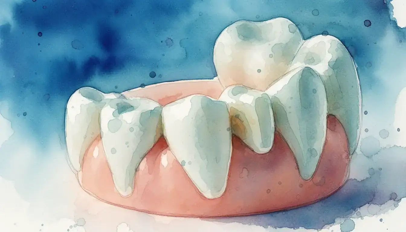
What Is Tooth Resorption?
Tooth resorption is a process where the body breaks down and absorbs its own tooth structure. Specialized cells dissolve the mineralized tissue of either the root or the internal chamber of a tooth. [1]
In a healthy mouth, a protective layer called cementum covers the root surface, and a layer of predentin lines the inner pulp chamber. These layers act as shields that prevent resorption from starting. When injury, infection, or other triggers damage these protective layers, the body may begin to treat the tooth like a foreign object and start dissolving it. [6]
Two main cell types drive this process. Odontoclasts break down dentin (the hard tissue that makes up most of the tooth). Osteoclasts break down cementum and bone. Both cell types are part of the body's normal remodeling system, but in resorption they become activated against the tooth itself. [1]
Tooth resorption is broadly divided into two categories based on where the breakdown begins. Internal resorption starts inside the pulp chamber or root canal. External resorption starts on the outer surface of the root. Each type has distinct causes, imaging characteristics, and treatment approaches. [4] Fuss et al. proposed a classification system based on the stimulating factors behind the resorption, which helps guide treatment decisions. [5]
Causes and Risk Factors
Tooth resorption is typically triggered by damage to the tooth's protective layers from trauma, infection, or other stimulating factors. [5]
The exact mechanism varies by type, but the underlying pattern is similar. Something disrupts the protective barriers on the root surface or inside the pulp. Inflammatory cells then move in, and the resorption process begins. Understanding these triggers is important because treatment often depends on identifying and removing the cause. [1]
Dental Trauma
Physical injury to a tooth is one of the most common triggers for resorption. A blow to the mouth can damage the cementum on the root surface or injure the pulp tissue inside the tooth. [1] Even if the tooth appears fine after an injury, microscopic damage to the root surface can set the stage for resorption months or years later.
Reimplantation of an avulsed (knocked-out) tooth carries a particularly high risk. When a tooth is knocked out and placed back in its socket, the root surface is often damaged. The longer the tooth stays outside the mouth, the greater the risk. Replacement resorption, a type where bone gradually replaces the root, is a well-known complication of tooth reimplantation. [5] [6]
Orthodontic Treatment
Orthodontic forces applied during braces or aligner treatment can trigger external root resorption. The pressure on the root surface during tooth movement creates areas of compression. In most cases the resorption is minor and stops when treatment ends. However, in some patients, the root shortening can be significant. [1]
Risk factors for orthodontic-related resorption include prolonged treatment time, heavy force application, and individual patient susceptibility. Teeth with a history of previous trauma are at higher risk during orthodontic treatment. [1]
Chronic Infection and Inflammation
Chronic pulp infection or periapical infection (infection at the tip of the root) can stimulate external resorption from the inside out. Bacteria and inflammatory byproducts damage the root surface. [5] Periodontal disease (gum disease) can also contribute by creating chronic inflammation around the root.
Impacted teeth, especially wisdom teeth that press against the roots of adjacent teeth, can cause pressure resorption. Cysts and tumors in the jawbone may also stimulate resorption as they expand and press against nearby roots. [1] [6]
Other Risk Factors
Some internal dental procedures can trigger resorption. Aggressive bleaching agents placed inside a tooth during internal whitening may seep through the dentin and damage the root surface, leading to cervical resorption. [1] Certain systemic conditions and hormonal factors have also been associated with increased susceptibility, though evidence on these links remains limited. [6]
Symptoms and Diagnosis
Most tooth resorption causes no pain or visible symptoms in its early stages. It is usually found by chance on a routine dental X-ray. [5]
What Patients May Experience
In the early phases, you will likely feel nothing at all. The tooth may look and function normally. This is what makes resorption particularly challenging; by the time symptoms appear, significant damage may already have occurred. [1]
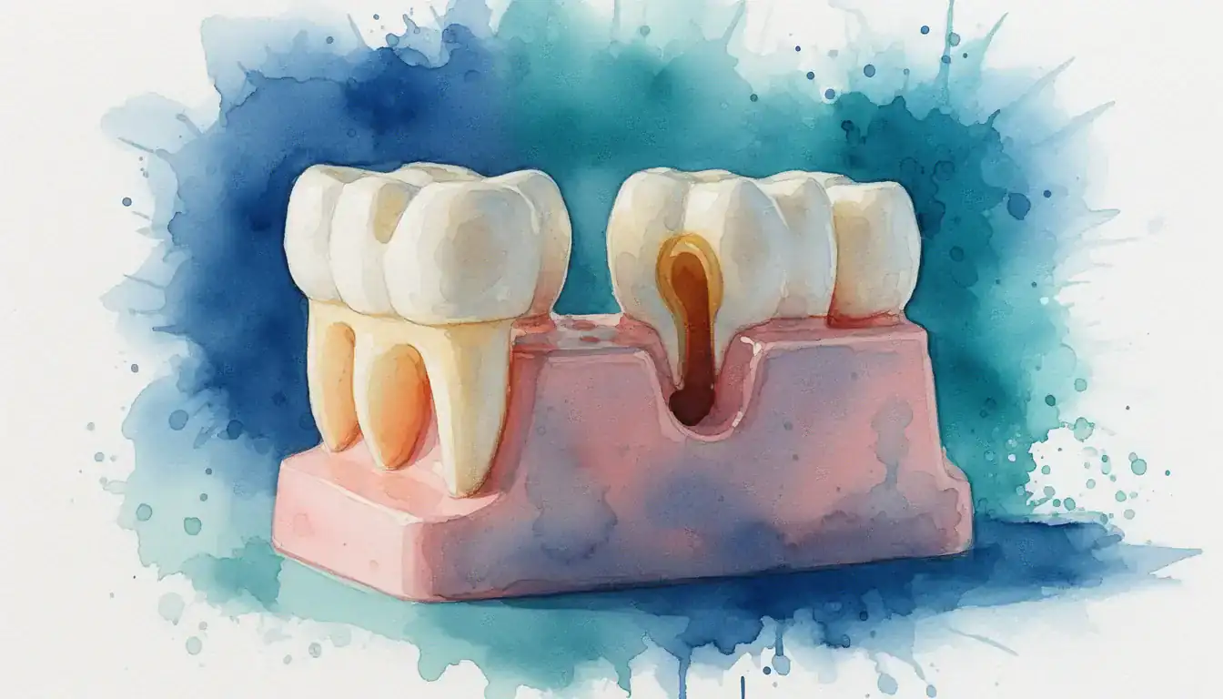
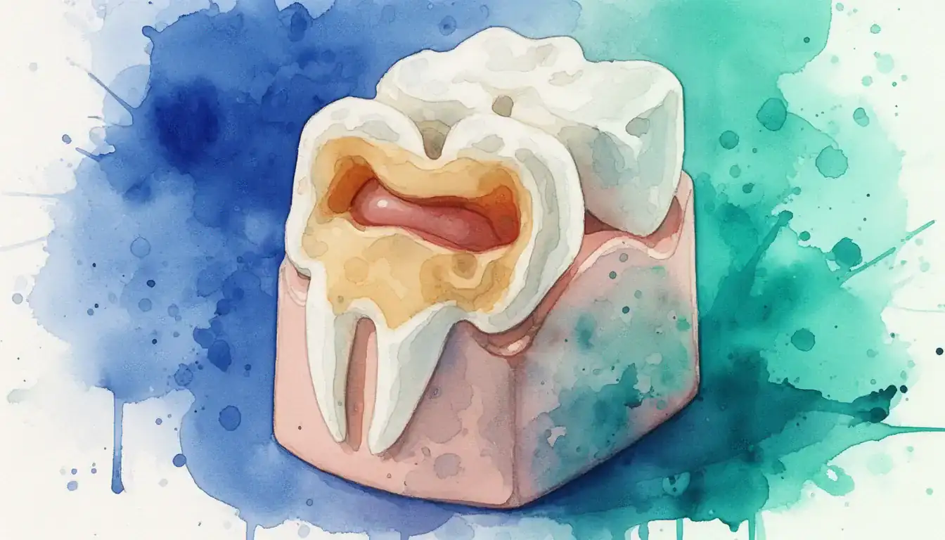
As resorption progresses, possible signs include a pinkish discoloration of the tooth crown (sometimes called a "pink tooth"), which happens when the resorbing tissue inside becomes visible through the thinning enamel. In advanced cases, there may be tooth mobility (looseness), dull pain, or swelling near the affected tooth. External cervical resorption near the gum line may sometimes look like a cavity at the base of the tooth. [4]
How Resorption Is Diagnosed
Standard periapical X-rays (small dental X-rays that show the whole tooth and root tip) are often the first tool used to detect resorption. On an X-ray, resorption typically appears as a dark area within or along the root. [4]
Distinguishing between internal and external resorption on a two-dimensional X-ray can be difficult. Patel et al. describe specific radiographic features that help differentiate the two types. Internal resorption often appears as a well-defined, round or oval dark area within the root canal. The outline of the root canal may seem to balloon outward. External resorption tends to have irregular margins and may overlap with the normal root canal outline. Shifting the X-ray angle can help: with internal resorption, the dark area stays centered on the root canal; with external resorption, the lesion shifts relative to the canal. [4]
Cone-beam computed tomography, commonly called CBCT, provides three-dimensional images and is increasingly used for complex cases. CBCT scans show the exact location, size, and extent of the resorption defect. This information is critical for treatment planning, especially when surgery is being considered. [1]
Clinical testing may include vitality tests (checking whether the nerve inside the tooth is alive) and periodontal probing (measuring the depth of gum pockets around the tooth). These tests help the specialist build a full picture of the tooth's condition.
Treatment Options
Treatment for tooth resorption depends on the type, location, and severity of the damage. Options range from root canal therapy to surgical repair to extraction. [5]
The goal of any treatment is to stop the resorption process, preserve as much tooth structure as possible, and restore the tooth to function. In some cases, monitoring without immediate intervention may be appropriate if the resorption is very small and stable. [1]
Root Canal Treatment for Internal Resorption
Internal resorption depends on a blood supply from the living pulp tissue. Removing the pulp through root canal treatment stops the resorption by eliminating the cells that are causing it. [5] [6]
During the procedure, the endodontist removes the inflamed or resorbing pulp tissue, disinfects the canal system, and fills the space with a biocompatible material. In areas where the canal has widened due to resorption, a material called mineral trioxide aggregate (MTA) or a bioceramic sealer may be used because these materials can adapt to irregular spaces and promote healing. [1]
Success rates for root canal treatment of internal resorption are generally favorable when the resorption has not perforated (broken through) the root wall. If a perforation exists, the prognosis depends on its size and location. Small perforations can often be sealed during treatment. Large perforations, especially those below the bone level, carry a less predictable outcome. [5]
Surgical Repair for External Resorption
External cervical resorption and some forms of external inflammatory resorption may require a surgical approach. The endodontist or oral surgeon exposes the affected area of the root, removes the resorptive tissue, and restores the defect with a filling material. [1]
For external inflammatory resorption caused by infection, root canal treatment may be the primary intervention, since removing the source of infection can halt the resorption process. When the resorption is on the root surface and accessible only from outside the tooth, a surgical flap is raised to reach the defect. The diseased tissue is carefully removed, and the area is sealed. [5]
In cases of replacement resorption (ankylosis), where bone is gradually replacing the root, treatment options are more limited. There is currently no reliable method to stop replacement resorption once it has started. Monitoring the tooth and planning for eventual replacement with an implant or bridge is often the most practical approach. [6]
Extraction
When resorption has destroyed too much tooth structure, the tooth may not be salvageable. Extraction becomes the recommended option when the root is severely weakened or when the resorption defect extends so deeply that repair is not feasible. [5]
After extraction, the missing tooth can typically be replaced with a dental implant, bridge, or removable partial denture. Your specialist can discuss which replacement option best suits your situation. Timing of the replacement depends on the condition of the surrounding bone and soft tissue.
Monitoring and Observation
Small, stable areas of external resorption that are not progressing may be monitored with periodic X-rays rather than treated immediately. This is sometimes the case with minor root shortening discovered after orthodontic treatment. [1] If follow-up imaging shows the resorption is advancing, active treatment can then be initiated.
Recovery and Aftercare
Recovery depends on the type of treatment performed. Root canal treatment typically involves minimal downtime, while surgical repair may require a few days of healing.
After root canal treatment for internal resorption, mild soreness around the treated tooth is common for a few days. Over-the-counter pain relievers are usually sufficient. The tooth will need a permanent restoration, often a crown, to protect it from fracture. Your dentist will typically place the crown within a few weeks of completing the root canal. [7]
Surgical repair of external resorption may involve some swelling and discomfort in the days following the procedure. Your specialist will provide specific post-operative instructions, which typically include soft foods for a few days, gentle oral hygiene around the surgical site, and possibly a short course of prescribed medication.
Regardless of the treatment type, follow-up imaging is essential. Resorption can recur, and regular X-rays or CBCT scans allow your specialist to catch any changes early. Follow-up appointments are typically scheduled at 3 months, 6 months, and then annually, though the exact schedule varies by case. [5]
Cost Factors
The cost of treating tooth resorption varies widely depending on the type of treatment, the complexity of the case, and your geographic location.
Root canal treatment for a tooth with internal resorption typically ranges from $800 to $1,500 for a single-rooted tooth and $1,000 to $2,000 or more for a multi-rooted tooth. These figures do not include the cost of a crown, which may add $1,000 to $2,000. Costs vary by location, provider, and case complexity.
Surgical treatment for external resorption, such as an apicoectomy (surgical removal of the root tip) or surgical repair of a cervical defect, typically ranges from $900 to $2,500 per tooth. Again, costs vary by location, provider, and case complexity.
Dental insurance may cover a portion of endodontic treatment, but coverage varies significantly by plan. Root canal treatment is generally classified as a major procedure, and many plans cover 50% to 80% after the deductible is met. Surgical procedures may have different coverage levels. Contact your insurance provider to confirm your specific benefits before treatment.
Many endodontic offices offer payment plans or accept third-party financing options to help spread the cost over time. Ask about these options during your consultation.
When to See a Specialist
A general dentist can detect resorption on a routine X-ray. An endodontist is the specialist trained to determine the exact type and plan the best treatment. [7]
Endodontists complete two to three years of additional training beyond dental school, focused specifically on conditions inside the tooth and around the root. They use operating microscopes, CBCT imaging, and microsurgical instruments that allow them to work with precision on complex resorption cases. [7]
You should ask your dentist about a referral to an endodontist if a routine X-ray shows a suspicious dark area in or around a root, if a tooth changes color (especially a pink or dark discoloration), if a tooth becomes loose without an obvious cause, or if you have a history of dental trauma and notice any changes in a previously injured tooth.
Early referral typically leads to more treatment options and better outcomes. A tooth with a small resorption defect is much more likely to be saved than one with extensive damage. Your general dentist and endodontist will work together to coordinate your care. Visit the endodontics page to learn more about what these specialists do.
Find an Endodontist Near You
If you or your dentist suspect tooth resorption, connecting with an endodontist is a smart next step. Use the My Specialty Dentist directory to search for endodontists in your area who have experience diagnosing and treating all types of tooth resorption. You can filter by location, read about each provider's background, and request a consultation directly through their profile.
Search Endodontists in Your Area