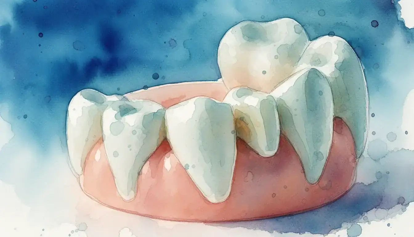
What Is a Periapical Lesion?
A periapical lesion is a pocket of inflamed or infected tissue that forms in the bone surrounding the tip of a tooth root.[8] It develops when bacteria or their byproducts escape from inside the tooth and trigger the body's immune response in the nearby bone.
Periapical lesions are among the most common findings on dental X-rays. Research shows that the majority of these lesions appear on teeth that have lost pulp vitality (the nerve and blood supply inside the tooth has died).[4] Most lesions are inflammatory in nature rather than tumors, and they are usually classified as periapical granulomas, radicular cysts, or abscesses based on tissue analysis.[8]
The condition can stay silent for months or years. Patients often discover a lesion only when a dentist takes a routine X-ray or when symptoms like swelling or pain finally appear. Endodontists, dental specialists who focus on the inside of the tooth, are typically the providers who diagnose and treat these lesions. Learn more on the endodontics page.
Causes and Risk Factors
Periapical lesions almost always start with bacteria reaching the inside of the tooth, where they trigger inflammation that spreads to the surrounding bone.[8] Several local and whole-body factors can raise your risk.
Whole-Body Risk Factors
Systemic health affects how the body fights infection at the root tip. Research links uncontrolled diabetes, immune-suppressing conditions, and certain medications to higher rates of apical periodontitis and slower healing after treatment.[5] Patients with diabetes show higher rates of periapical inflammation, especially when blood sugar is poorly controlled.[9]
Patients taking antiresorptive drugs (such as bisphosphonates for osteoporosis) need extra planning before any endodontic surgery, because these medications can affect jawbone healing.[2] Genetics and individual immune response also help explain why some people develop lesions while others with similar dental issues do not.[6]
Symptoms and Diagnosis
Many periapical lesions cause no symptoms at all. When symptoms appear, they range from mild discomfort to severe pain, and a clinical exam plus X-rays confirm the diagnosis.[4][8]
What Patients Experience
Symptoms can include a dull ache, sensitivity to biting, swelling of the gum near the tooth, or a small bump that drains pus (called a sinus tract). Some patients notice a bad taste, mild fever, or tenderness when chewing.
Studies of vital versus nonvital teeth show that nonvital teeth (no living pulp) more often present with painless, longer-standing lesions, while vital teeth with apical periodontitis are more likely to cause sharp or throbbing pain.[4] Acute abscesses can spread quickly and cause facial swelling, which is a dental emergency.
How Endodontists Diagnose
Diagnosis combines several tests. The endodontist asks about your symptoms, taps on the tooth (percussion test), checks for swelling, and uses cold or electric pulp testing to check whether the nerve is alive.[8]
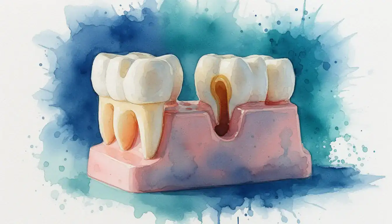
Imaging is essential. A periapical X-ray often shows the lesion as a dark area at the root tip. Cone-beam computed tomography (CBCT), a 3D dental scan, may be used for complex cases because it shows the lesion's true size and any nearby anatomy.[8] In rare cases, a biopsy is needed to rule out non-inflammatory lesions.
- Persistent toothache or sensitivity to pressure
- Swelling or a pimple-like bump on the gum
- Bad taste or foul drainage from the gum
- A discolored tooth that has darkened over time
- Facial swelling, fever, or trouble swallowing — seek same-day care
Treatment Options
The goal of treatment is to remove the source of infection so the bone can heal. Most lesions resolve with nonsurgical root canal treatment, but surgery, retreatment, or extraction are options when needed.[3][8]
Root Canal Treatment (First-Line)
Nonsurgical root canal treatment is usually the first option. The endodontist removes infected pulp tissue, disinfects the canal system, and seals it with a rubber-like material. Healing of the bone follows over 6 to 24 months as the immune system clears the inflammation.
Long-term studies report healing rates of 74% to 86% for primary root canal treatment of teeth with periapical lesions.[3] Healing depends on cleaning the canal thoroughly and sealing it well. Most teeth then need a crown to protect them from fracture.
Endodontic Retreatment
If a previous root canal has not healed, retreatment may be considered. The endodontist removes the old filling material, disinfects the canals again, and re-seals them. Retreatment success is generally lower than first-time treatment, but in many cases, it remains a tooth-saving option before surgery.[3]
Apical Surgery (Apicoectomy)
When retreatment is not possible or has already failed, the endodontist may recommend an apicoectomy. In this microsurgery, the tip of the root is removed along with the lesion, and the end of the canal is sealed from the outside. Modern microsurgical techniques have improved outcomes, with reported success rates often above 80% for well-selected cases.[3]
Regenerative Endodontics
For young patients with immature roots and a non-vital pulp, regenerative endodontic procedures may be an option. These techniques aim to encourage the body to rebuild tissue inside the canal so the root can continue to develop. A systematic review found favorable outcomes in selected cases, though indications remain specific.[7]
Extraction
If the tooth is cracked deep below the gum, has lost too much structure, or has not responded to other treatments, extraction may be the best option. After healing, replacement options include a dental implant, a bridge, or a partial denture. Each option has its own benefits and tradeoffs that an endodontist or general dentist can review with you.
Recovery and Aftercare
Recovery from root canal treatment is usually quick, with most patients returning to normal activity within a day or two. Bone healing of the lesion itself takes much longer and is tracked with follow-up X-rays.[8]
After a root canal, mild soreness for several days is normal and typically responds to over-the-counter pain relievers. Avoid chewing on the treated tooth until the final restoration (often a crown) is placed. After apical surgery, expect some swelling and bruising for several days, and follow your specialist's instructions on rinsing, diet, and stitches.
Healing is often confirmed with X-rays at 6, 12, and sometimes 24 months. The lesion gradually shrinks as new bone fills in. Conditions that affect healing, such as diabetes or smoking, may extend recovery and influence outcomes.[5][9] Tell your endodontist about any health changes or new medications between visits.
Cost Factors and Insurance
Treatment costs depend on which tooth is involved, how complex the case is, and where you live. Costs vary by location, provider, and case complexity, so always confirm fees with your endodontist's office before scheduling.
Typical fee ranges in the United States are roughly $700 to $1,800 for nonsurgical root canal treatment, $900 to $2,000 for retreatment, and $1,000 to $2,500 for apical surgery. A crown or other final restoration is usually a separate cost. Many dental insurance plans cover endodontic care under their basic or major services category, often paying 50% to 80% after the deductible is met.
Most endodontic offices accept dental insurance, health savings accounts (HSAs), and offer financing through providers like CareCredit. Ask whether the office is in-network for your plan and request a written treatment plan that lists each procedure code and fee. The American Dental Association provides patient guidance on understanding dental benefits.[12]
When to See an Endodontist vs. a General Dentist
General dentists handle many routine root canals, but endodontists are dental specialists who complete an extra two or more years of training focused on the inside of the tooth and the surrounding bone. The American Association of Endodontists encourages referral for complex cases.[11]
Consider an endodontist if your tooth has a periapical lesion that has not responded to a previous root canal, the canals are curved or calcified, the tooth has unusual anatomy, or you have whole-body health concerns that complicate treatment. Endodontists also use surgical microscopes and advanced imaging that can improve outcomes in difficult cases.
If your symptoms are severe, including facial swelling, fever, or difficulty swallowing, seek care immediately. These can be signs of a spreading infection that needs urgent treatment, sometimes with antibiotics and drainage in addition to definitive endodontic care.
Find an Endodontist Near You
If a dentist has spotted a dark spot at the tip of your tooth root, or you have ongoing pain or swelling, an endodontist can review your X-rays, confirm the diagnosis, and walk you through your options. Visit the endodontics page to find a board-certified endodontist in your area and learn what to expect at your first visit.
Search Endodontists in Your Area